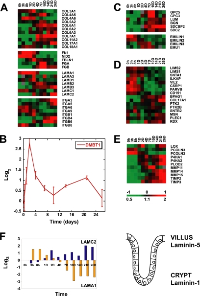
Figure 5.
Temporal expression patterns identified for genes encoding cell-ECM adhesion molecules during in vitro development of cell polarity, including family members of collagen, laminin and integrin receptors (A), hensin (DMBT1) (B), heparan sulfate proteoglycans (C), and hemidesmosome and focal adhesion complex components (D). Enzymes with roles in the modification or remodeling of ECM are also under fine transcriptional regulation during Caco-2 cell polarization (E). (F) Left, the reciprocal expression pattern observed for transcripts encoding components of laminin-1 and -5 (LAMA1 and LAMC2, respectively) during in vitro establishment of an epithelial layer, which mimic in vivo expression trends of laminin chains, previously identified along the human intestinal crypt villus axis (shown on the right; Leivo et al., 1996; Orian-Rousseau et al., 1996). Transcript levels determined by microarray analysis are shown relative to a reference pool of human mRNAs. A summary of the roles of individual cell–ECM components is listed in Supplementary Table S1.

