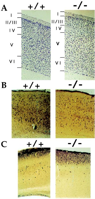
Figure 2.
Defects in the motor area of the neo-cortex of FGF2−/− mice. Coronal sections of the FGF2−/− mutant cortex compared with those of wild-type FGF2+/+ cortex after cresyl violet (NISSL) staining (A), parvalbumin (B), and calbindin (C) immunohistochemistry. Reduced cell density is evident in layers V and VI in A–C.

