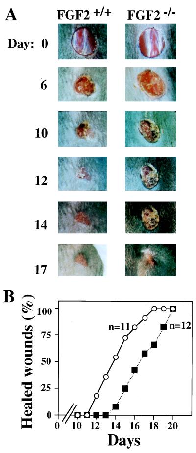
Figure 3.
Skin wound healing of FGF2−/− mice. (A) Example of the healing progress in an FGF2−/− mouse and an FGF+/+ mouse. Wounds were photographed at the time indicated. Day 0 picture was taken immediately after wounding. All wounds were photographed at the same distance. (B) The fraction of completely healed wounds (determined as described in Materials and Methods) is plotted versus the time after wounding. n, number of mice of each genotype analyzed. Wounds were performed in a genotype-blind fashion. Mice were caged separately throughout the experiment. ○, FGF2+/+; ▪, FGF2−/−.

