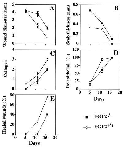
Figure 4.
Histologic analysis of wound healing in FGF2−/− and FGF2+/+ mice. Wound diameter (A), scab thickness (B), collagen deposition (C), percentage of reepithelialization (D), and percentage of completely healed wounds (E) are plotted versus time after wounding. The number of animals was five FGF2−/− and five FGF2+/+ at 5 days and 16 days after wounding, respectively, and seven FGF2−/− and seven FGF2+/+ at day 11. Each point represents the mean ± SEM, except in E. Wounds that were completely healed were not considered in the calculation of the mean. The histologic parameters were determined as described in Materials and Methods.

