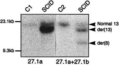
Figure 3.
FIM rearrangement in t(8;13) cells. Southern blot analysis of BamHI-digested DNAs from SCID t(8;13) and controls (C1, mammary carcinoma MDA-MB-134 cells; C2, normal lymphocytes). Hybridizations were done with the FIM cDNA 27.1a probe (two first lanes) and 27.1a plus 27.1b. Positions of the normal and the two rearranged fragments are indicated (arrowheads) on the right. The sizes of HindIII-digested λ DNA fragments are indicated on the left.

