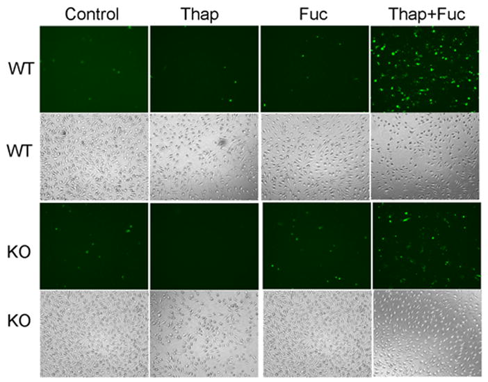
FIGURE 4. Fluorescence microscopic images of apoptosis induced by incubating peritoneal macrophages from wild-type and iPLA2β-null mice with thapsigargin and fucoidan.

The leftmost column (1st) represents control, untreated peritoneal macrophages from wild-type (WT) or iPLA2β-knock-out (KO) mice, and the other columns represent macrophages incubated (24 h) with thapsigargin alone (Thap), fucoidan alone (Fuc), or both (Thap + Fuc). In the fluorescence micrographs in the 1st (topmost) and 3rd rows, externalization of phosphatidylserine was visualized with Alexa-488-labeled annexin V. Light micrographs in the 2nd and 4rth rows display total cells.
