
FIGURE 7. Cytochrome c release into cytosol of wild-type and iPLA2β-null mouse macrophages incubated with thapsigargin and fucoidan.
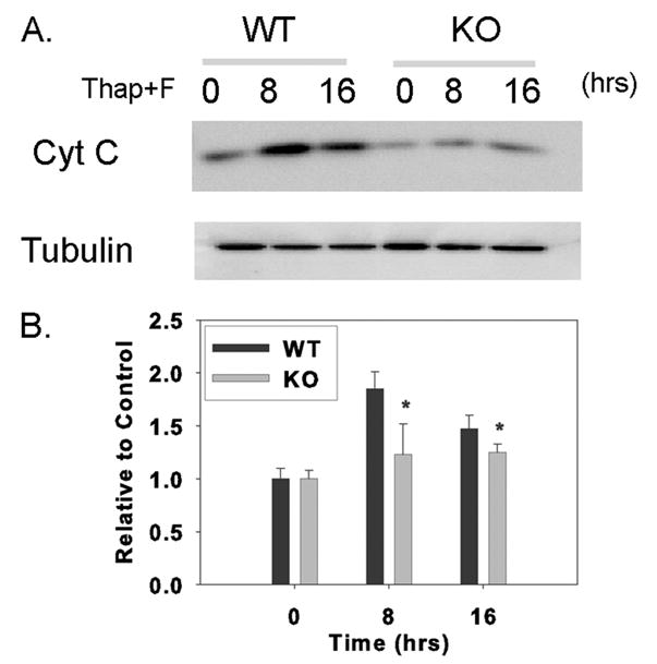
Wild-type (WT) or iPLA2β-knock-out (KO) macrophages were incubated without or with thapsigargin and fucoidan (Thap + F) for various intervals. At the end of the incubations, cytosol was prepared, analyzed by SDS-PAGE, electrotransferred to nylon membranes, and probed with cytochrome c and tubulin antibodies, as illustrated in A. B represents densitometric ratios of iPLA2β and tubulin immunoblot signals determined with AlphaEaseFC software. Mean values ± S.E. are displayed (n = 4). Asterisk denotes p < 0.05 for WT versus KO.