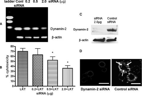
FIG. 5.
siRNA knockdown of dynamin-2 reduces LKT-mediated cytotoxicity and internalization in BL-3 cells. Dynamin-2 was knocked down by transfecting BL-3 cells with siRNA (0.2, 0.5, or 2.0 μg) for 6 h, followed by further incubation in RPMI medium with 10% fetal bovine serum for 72 h. Control cells were transfected with a scrambled siRNA sequence for the same time period. To assess the degree of dynamin-2 RNA knockdown, RNA was extracted, and a one-step reverse transcription-PCR assay with dynamin-2-specific primers was performed. In panel A the products were visualized by electrophoresis in a 1% agarose gel. In panel B, dynamin-2 siRNA- or scrambled siRNA-transfected BL-3 cells (106) were incubated with LKT (0.5 U) for 1 h, and cytotoxicity was measured as described previously. The data are the means and standard errors of the means of five separate siRNA transfection experiments. An asterisk indicates that the P value is <0.05 for a comparison with the scrambled siRNA control. Panel C shows an immunoblot analysis of dynamin-2 knockdown from BL-3 cells transfected with 2 μg of dynamin-2 siRNA. In panel D, BL-3 cells transfected with dynamin-2 siRNA (2.0 μg) or with scrambled siRNA were fixed, permeabilized with cold acetone, blocked with 3% bovine serum albumin, and incubated with anti-LKT MAb. A goat anti-mouse IgG-Texas Red-conjugated secondary antibody was added, and the LKT signal was visualized by fluorescent microscopy at excitation and emission wavelengths of 595 and 614 nm, respectively. The arrows indicate internalized LKT. Bars = 10 μm. Conl, control.

