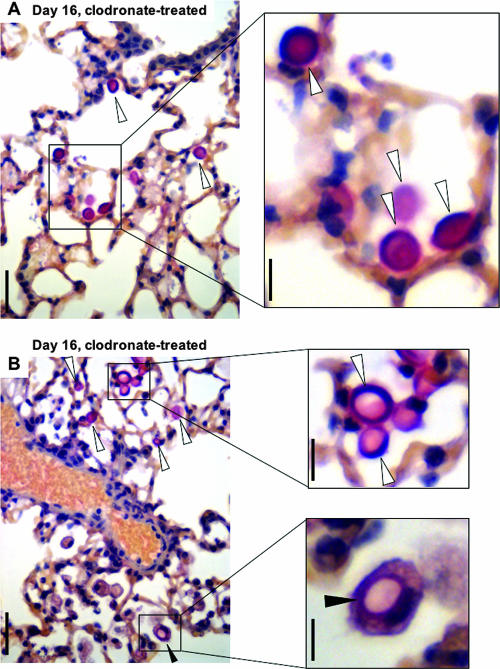
FIG. 6.
Histopathology of mucicarmine-stained Tgɛ26 lungs infected with the Δgcs1 strain and treated with clodronate. Panels A and B show lung fields from two different mice at day 16 of infection. The open arrowheads indicate extracellular Δgcs1 cells. The filled arrowheads in panel B indicate a Δgcs1 cell within a macrophage. Bars = 50 μm (left panels) and 10 μm (right panels).

