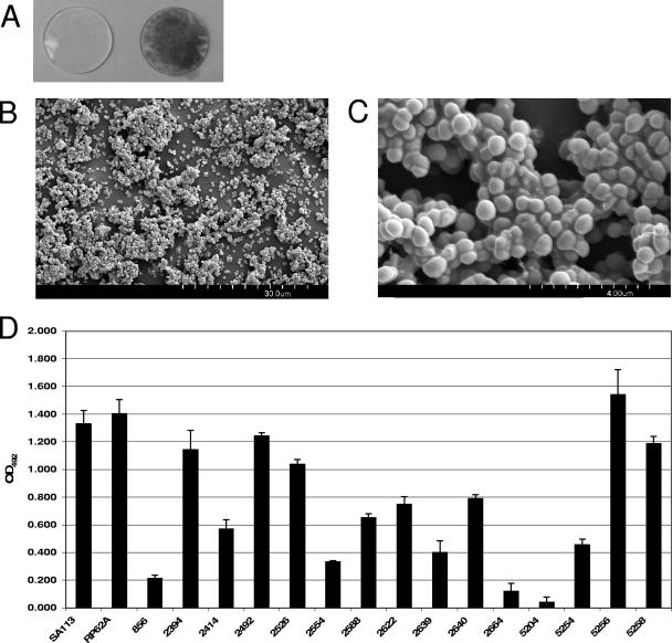
FIG. 1.
Characterization of biofilm formation by S. lugdunensis clinical isolates. (A) S. lugdunensis biofilm formation on silicone elastomer. Disks cut from nonreinforced silicone elastomer sheeting were sterilized and incubated for 24 h in TSBgluc1% (left) or TSBgluc1% containing S. lugdunensis IDRL-2640 (right). Disks were rinsed and stained with safranin to visualize biofilms. (B) Scanning electron micrograph (magnification, ×1,800) of S. lugdunensis IDRL-2640 biofilm formed on a silicone elastomer disk after 24 h. (C) Higher-magnification (×13,000) SEM of biofilm shown in panel B. (D) Biofilm formation by S. aureus SA113, S. epidermidis RP62A, and 15 clinical S. lugdunensis isolates on polystyrene when grown in TSBgluc1% for 24 h. Biofilms were stained with safranin, resuspended in 30% glacial acetic acid, and quantified by OD492. Bars are the averages of measurements taken from four duplicate wells. Error bars represent the standard deviations. The assay was repeated three times, and representative data from a single replicate are shown.

