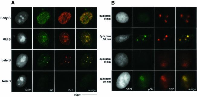
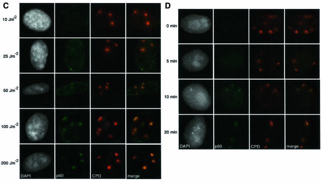
Fig. 2. Recruitment of CAF-1 p60 to replication and damage sites. (A) Characteristic patterns of CAF-1 p60 staining throughout the cell cycle. Cells were pulsed with BrdU followed by double labelling with a polyclonal against p60 (green) and a rat monoclonal against BrdU after denaturation with 4 M HCl (red). (B) CAF-1 p60 is locally recruited to sites of UV damage. Cells were irradiated with 100 J/m2 through filters with either 3 µm or 8 µm pores, with or without post-irradiation incubation, as indicated. Indirect immunofluorescence was performed with a polyclonal against p60 (green) and the anti-thymine dimer mouse monoclonal antibody (red). (C) Damage sites and CAF-1 p60 were visualized as in (B), 30 min after irradiation at the doses indicated. (D) Damage sites and CAF-1 p60 were visualized as in (B), at different times after a dose of 150 J/m2.

An official website of the United States government
Here's how you know
Official websites use .gov
A
.gov website belongs to an official
government organization in the United States.
Secure .gov websites use HTTPS
A lock (
) or https:// means you've safely
connected to the .gov website. Share sensitive
information only on official, secure websites.

