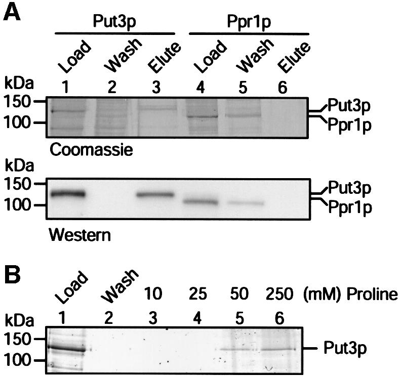
Fig. 6. Direct interaction between Put3p and proline. (A) Samples of purified Put3p or Ppr1p were applied to a l-proline–EAH Sepharose column. The flow-through from each column (wash) and the material eluted in the presence of 250 mM l-proline were separated by SDS–PAGE and either stained with Coomassie Blue (top) or visualized as a western blot to detect the polyhistidine tag of the protein (bottom). The sizes of molecular weight standards (in kDa) are indicated. (B) Elution of Put3p from a l-proline–EAH Sepharose column using different concentrations of l-proline. Samples of purified protein were applied, in parallel, to proline-affinity columns. Unbound proteins were washed from the column and then bound protein eluted using l-proline at the concentration indicated. Samples were separated by SDS–PAGE and subjected to silver staining.
