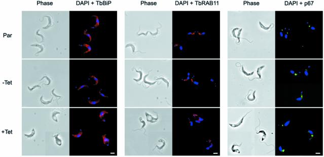
Fig. 4. Preservation of normal morphology of internal membrane structures in BigEye cells. Immunofluorescence analysis for endoplasmic reticulum (TbBiP), recycling endosome (TbRAB11) and lysosomal (p67) proteins. Top, the parental BSF 90-13 cells (Par); middle, uninduced bloodstream form (BSF) RNAi line (–Tet); and bottom, induced (+Tet). Induction is confirmed by the presence of the BigEye. The distribution of marker proteins is unaltered between cell lines. Left, phase; right, DAPI (DNA) in blue, TbBiP or TbRAB11 in red, p67 in green. Scale bar: 2 µm.

An official website of the United States government
Here's how you know
Official websites use .gov
A
.gov website belongs to an official
government organization in the United States.
Secure .gov websites use HTTPS
A lock (
) or https:// means you've safely
connected to the .gov website. Share sensitive
information only on official, secure websites.
