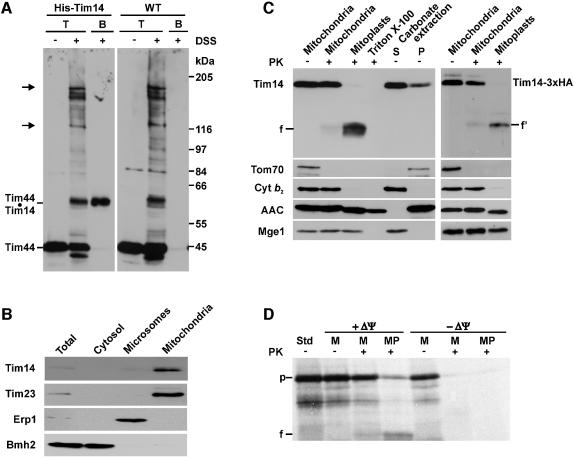
Fig. 2. Tim14 is a component of the inner membrane of mitochondria. (A) Tim14 is the product of the YLR008c reading frame. Mitochondria isolated from a yeast strain in which a sequence encoding eight histidine residues was fused to the 5′ end of the YLR008c reading frame, and from wild-type cells, were subjected to crosslinking with DSS. An aliquot of each type of mitochondria was directly subjected to SDS–PAGE; the other aliquot was solubilized and incubated with Ni-NTA beads. Bound material was eluted and subjected to SDS–PAGE. Resolved proteins were blotted onto nitrocellulose membrane and immunodecorated with antibodies against Tim44. T, total mitochondria incubated in the absence or presence of DSS; B, material bound to Ni-NTA beads. Arrows indicate crosslinked adducts of Tim44 to mtHsp70. (B) Tim14 is located in mitochondria. Equal amounts of protein of subcellular fraction were subjected to SDS–PAGE and immunodecoration with antibodies against Tim14 and marker proteins of mitochondria (Tim23), microsomes (Erp1) and cytosol (Bmh2). (C) Tim14 is located in the inner membrane, exposing an N-terminal segment of ∼8 kDa into the intermembrane space and its C-terminus into the matrix. Left panel: isolated mitochondria, mitoplasts prepared by osmotic shock, Triton-solubilized mitochondria and the supernatant (S) and pellet (P) of carbonate extraction were incubated with or without proteinase K (PK; 100 µg/ml). Samples were subjected to SDS–PAGE and immunoblotting with antibodies against Tim14 and against various mitochondrial marker proteins. Cytb2, cytochrome b2; AAC, ADP/ATP carrier; f, 13 kDa fragment of Tim14. Right panel: mitochondria containing a version of Tim14 carrying a 3HA-tag at its C terminus and derived mitoplasts were treated with proteinase K as indicated. Samples were analyzed by SDS–PAGE and immunoblotting with an antibody against the HA-tag and against the indicated mitochondrial marker proteins. f′, fragment. (D) Tim14 can be imported into isolated mitochondria. Reticulocyte lysate containing 35S-labeled Tim14 was incubated with mitochondria in the presence or absence of a membrane potential ΔΨ. Mitochondria were reisolated, aliquots were converted to mitoplasts and treated with proteinase K. Samples were subjected to SDS–PAGE and autoradiography. M, mitochondria; MP, mitoplasts; Std, 40% of input into import experiments; p, precursor of Tim14; f, 13 kDa fragment.

An official website of the United States government
Here's how you know
Official websites use .gov
A
.gov website belongs to an official
government organization in the United States.
Secure .gov websites use HTTPS
A lock (
) or https:// means you've safely
connected to the .gov website. Share sensitive
information only on official, secure websites.
