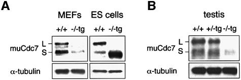
Fig. 7. Diminished expression of Cdc7 in Cdc7–/–tg MEFs and testes. The levels of Cdc7 protein in MEFs [(A), left], ES cells [(A), right] and testes (B) were determined by western blot analysis. Cdc7–/–tg MEFs express Cdc7 protein at levels five times lower than that in wild-type MEFs, whereas the level of Cdc7 in Cdc7–/–tg ES cells is ∼3- to 5-fold higher than that of the wild-type ES cells. The Cdc7 protein level in Cdc7–/–tg testes is 10 times lower than those in the wild-type and Cdc7+/–tg testes. α-tubulin is shown as a loading control. L and S denote two alternative spliced forms of murine Cdc7 protein, which are identical in functions. The transgene encodes the form S.

An official website of the United States government
Here's how you know
Official websites use .gov
A
.gov website belongs to an official
government organization in the United States.
Secure .gov websites use HTTPS
A lock (
) or https:// means you've safely
connected to the .gov website. Share sensitive
information only on official, secure websites.
