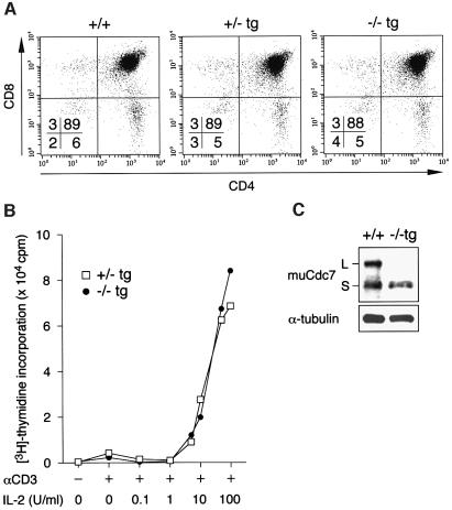
Fig. 8. Thymic development was normal in Cdc7–/–tg mice. (A) Flow-cytometric analysis of thymocytes from Cdc7–/–tg mice. Expression of CD4 and CD8 in thymocytes of Cdc7–/–tg mice and their control littermates was analyzed. Proportions of cells in the quadrants are shown. Staining and analysis were performed on at least three animals of each genotype with similar results. (B) Thymocytes prepared from Cdc7+/–tg and Cdc7–/–tg mice were stimulated using anti-CD3ε antibody and IL-2 of various doses. Mitogenic activation of Cdc7–/–tg thymocytes is unperturbed. Data shown are averages of three independent experiments. (C) The levels of Cdc7 protein in thymi from wild-type (+/+) and Cdc7–/–tg mice were determined by western blot analysis with anti-murine Cdc7 antibody. Cdc7–/–tg thymi express Cdc7 protein at the level ∼65% that of wild-type thymi. α-tubulin is shown as a loading control. L and S denote two alternative spliced forms of murine Cdc7 protein.

An official website of the United States government
Here's how you know
Official websites use .gov
A
.gov website belongs to an official
government organization in the United States.
Secure .gov websites use HTTPS
A lock (
) or https:// means you've safely
connected to the .gov website. Share sensitive
information only on official, secure websites.
