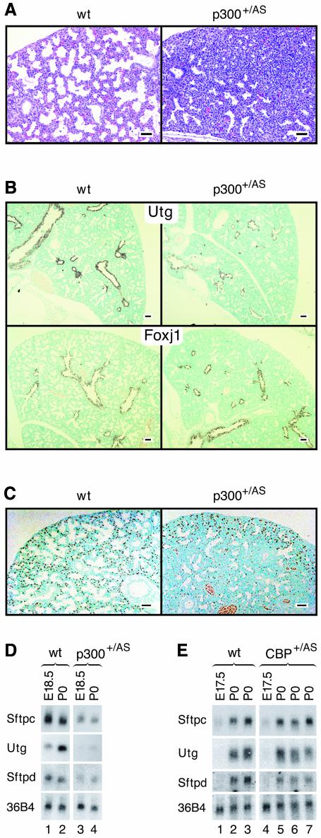
Fig. 3. Defective saccule formation and impaired proximal as well as distal epithelial cell differentiation in p300+/AS lungs. (A) HE staining of lung sections from wild-type and p300+/AS embryos at E18.5, showing condensed tissue with little evidence for saccule formation in the mutant. (B) In situ hybridization for proximal markers Utg (top panels) and Foxj1 (bottom panels). (C) Immunohistochemistry for the distal endoderm marker protein surfactant protein C showing that positive cells are confined to the periphery of p300+/AS mutant lungs. Two blood vessels are visible near the bottom part of the right panel. (D and E) Northern blot analysis of the indicated epithelial marker transcripts in p300+/AS and Cbp+/AS lungs. 36B4 served as internal standard. Bars = 100 µm.

An official website of the United States government
Here's how you know
Official websites use .gov
A
.gov website belongs to an official
government organization in the United States.
Secure .gov websites use HTTPS
A lock (
) or https:// means you've safely
connected to the .gov website. Share sensitive
information only on official, secure websites.
