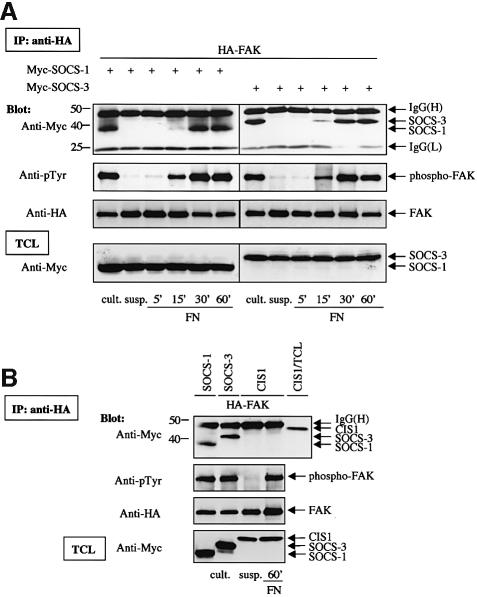
Fig. 2. Adhesion-dependent interaction of SOCS-1 and SOCS-3 with FAK. (A) COS-7 cells were transiently transfected with the indicated plasmids (0.2 µg of HA-FAK, 0.5 µg of Myc-SOCS-1/3). Twenty-four hours after transfection, cells were serum-starved for additional 24 h, detached, kept in suspension for 30 min (‘susp.’), and replated or not on FN for the indicated times. Alternatively, cells were lysed as a monolayer culture 48 h after transfection without prior additional manipulation (‘cult.’). Cell lysates were immunoprecipitated with anti-HA antibody, and the precipitates were analyzed by immunoblotting with antibodies against Myc, phosphotyrosine (pTyr) or HA. Total cell lysates were subjected to immunoblotting with anti-Myc antibodies to confirm SOCS-protein expression levels. (B) COS-7 cells were transiently transfected with the indicated plasmids (0.2 µg of HA-FAK, 0.5 µg of Myc-SOCS-1/3 or CIS1). Twenty-four hours after transfection, CIS1-FAK-cotransfected cells were serum-starved for additional 24 h, detached, kept in suspension for 30 min, and replated or not on FN for 1 h. FAK-SOCS-1/3-cotransfected cells were treated as described above for monolayer culture. Cell lysates were immunoprecipitated with anti-HA antibody, and the precipitates were analyzed by immunoblotting with antibodies against Myc, pTyr or HA. An aliquot of total cell lysate from CIS1-transfected cells was included as a control in the anti-Myc immunoblot of anti-HA immunoprecipitates to determine the motility of the CIS1 protein relative to the IgG band. Total cell lysates of all samples were subjected to immunoblot with anti-Myc antibody to confirm SOCS-protein expression levels.

An official website of the United States government
Here's how you know
Official websites use .gov
A
.gov website belongs to an official
government organization in the United States.
Secure .gov websites use HTTPS
A lock (
) or https:// means you've safely
connected to the .gov website. Share sensitive
information only on official, secure websites.
