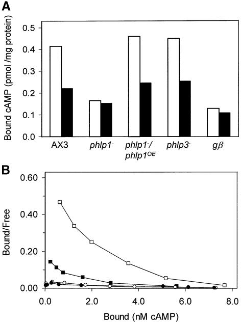
Fig. 4. Defective receptor–G protein interaction in phlp1– cells. (A) GTPγS inhibition of 3H-cAMP binding to membranes of AX3, phlp1–, phlp1–/phlp1OE and gβ– cells. Membranes were prepared from cells that were starved for 5 h. Binding assays were performed in the absence (open bars) or presence (black bars) of 30 µM GTPγS. (B) To determine the number and affinity of the cAMP-binding sites, Scatchard analysis was carried out by including different concentrations of cAMP in the binding assays. The results for membranes of phlp1–/phlp1OE (squares) or phlp1– cells (circles) in the absence (open symbols) or presence (closed symbols) of 30 µM GTPγS are shown. One nanomole of bound cAMP is equivalent to 6000 binding sites per cell. Data were fitted using the program FigP. The data for phlp1–/phlp1OE in the absence of GTPγS were fitted with a two-receptor model; the data for the other conditions were fitted statistically better with a one-receptor model. The kinetic data are as follows: for phlp1–/phlp1OE without GTPγS, Kd1 = 4.07 ± 3.68 nM, B1d = 15 600 ± 2600 sites/cell, Kd2 = 557 ± 107 nM and B2 = 84 000 ± 17 000 sites/cell; for phlp1–/phlp1OE with GTPγS, Kd = 507 ± 92 nM and B = 80 500 ± 6000 sites/cell; for phlp1– without GTPγS, Kd = 480 ± 35 nM and B = 60 000 ± 2500 sites/cell; for phlp1– with GTPγS, Kd = 491 ± 31 nM and B = 65 000 ± 2000 sites/cell.

An official website of the United States government
Here's how you know
Official websites use .gov
A
.gov website belongs to an official
government organization in the United States.
Secure .gov websites use HTTPS
A lock (
) or https:// means you've safely
connected to the .gov website. Share sensitive
information only on official, secure websites.
