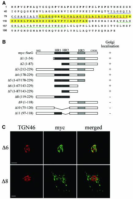
Fig. 6. Identification of Golgi-targeting regions of SseG. (A) Amino acid sequence of SseG. Hydrophobic regions HR1, HR2 and HR3 are underlined in blue, red and green, respectively. The Golgi targeting region is highlighted in yellow. Prolines in the N-terminal 31 amino acids are in bold. (B) Schematic representation of truncated polypeptides derived from myc::SseG. The extent of each deletion is shown in parentheses to the left of each construct. The ability of each polypeptide to associate with the Golgi is indicated to the right of each construct. (C) Representative images of Δ6 (upper panel) showing extensive co-localization with TGN46 and Δ8 (lower panel) showing a scattered distribution, with no co-localization with the Golgi marker. Scale bars correspond to 5 µm.

An official website of the United States government
Here's how you know
Official websites use .gov
A
.gov website belongs to an official
government organization in the United States.
Secure .gov websites use HTTPS
A lock (
) or https:// means you've safely
connected to the .gov website. Share sensitive
information only on official, secure websites.
