
Figure 3.
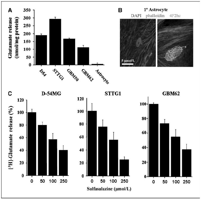
Glutamate release by glioma cells is inhibited by sulfasalazine in a dose-dependent manner. A, glutamate released by four glioma cell lines were compared with primary cultured astrocytes using an enzyme-based bioluminescent signal normalized to protein. B, images of primary cultures of rat astrocytes show a control (left) and positive staining for 4F2hc on the right, labeling only the Golgi, in green. DAPI-labeled nuclei and phalloidin (a cytoskeleton protein conjugated with Alexa 546) are included. System is nonfunctional in astrocyte cultures. C, glutamate release is shown in the presence of increasing concentrations of sulfasalazine using [3H]glutamate-loaded glioma cell lines to measure release. Columns, percent control; bars, SE. All experiments were done with at least n = 3.