
Figure 5.
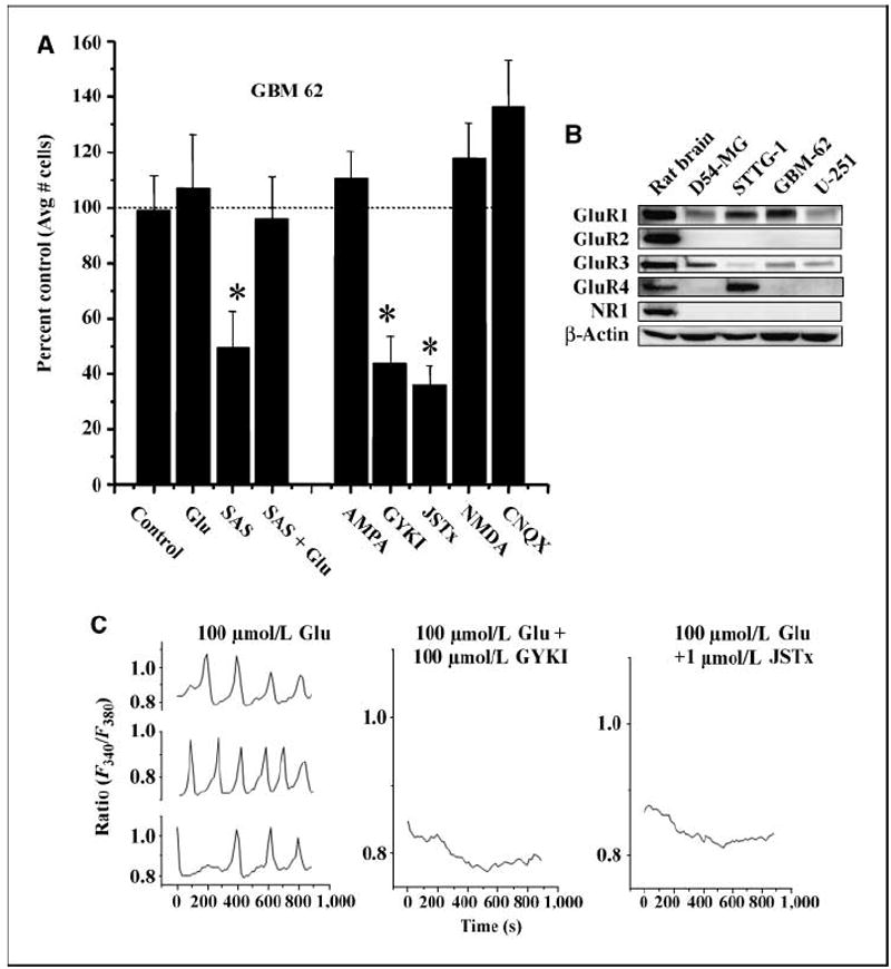
Glutamate induces AMPA-R–dependent calcium oscillations in glioma cells. A, the invasion results of patient-derived glioma cells (GBM 62) are shown in the presence of sulfasalazine (250 μmol/L SAS) and SAS + glutamate (100 μmol/L). Using sister wells, in the second half of A, glioma cells were allowed to invade the filter pores in the presence of the AMPA-R agonist AMPA (100 μmol/L); AMPA-R antagonists GYKI (100 μmol/L) and Joro spider toxin (JSTx; 1 μmol/L); NMDA-R agonist NMDA (100 μmol/L); and AMPA/KA-R antagonist, CNQX (100 μmol/L). Each experiment had three similarly treated inserts where six fields per insert were imaged and data averaged. This was repeated thrice. Bars, SE * P ≤ 0.05. B, glioma cells express Ca2+-permeable AMPA-Rs as shown by Western blot analysis for four glioma cell lines with whole rat brain lysates as a positive control. Glutamate subunits of AMPA-Rs were probed with antibodies to GluR1-4. β-Actin is shown as a control for protein loading efficiency. C, representative traces from three glioma cells loaded with FURA2-AM showed oscillatory changes in [Ca2+]i in response to 100 μmol/L glutamate. Single representative traces are shown following simultaneous application of 100 μmol/L glutamate and the AMPA-R blockers GYKI (100 μmol/L) and Joro spider toxin showing that [Ca2+]i oscillations stimulated by glutamate are inhibited. Each experiment was repeated three independent times.