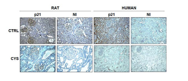
Figure 2.
Representative immunohistochemical expression of p21 in male cystic and noncystic Han:SPRD rats (left), and male PKD and control humans (right). Upper panels show p21 localization from control sections, and the lower panels show cystic sections. Also shown are sections (NI) pre-incubated with non-immune serum that demonstrate antibody specificity. In sections from control male kidneys, p21 was widely distributed, whereas in sections from cystic male kidneys, p21 was only sparsely detected.

