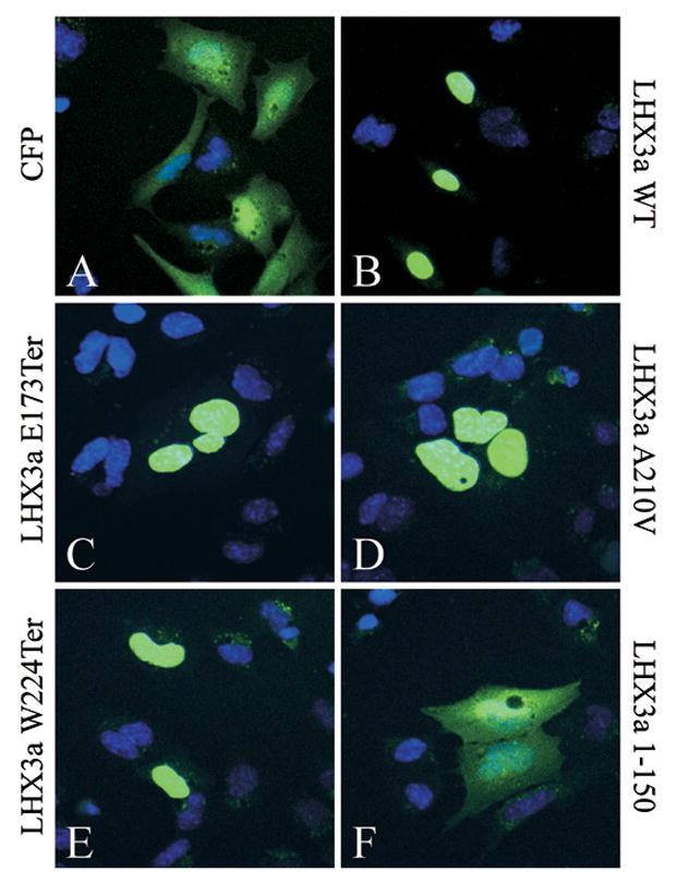
Fig. 6.

Intracellular localization of wild type (WT) and mutant LHX3a proteins fused to the cyan fluorescent protein (CFP). The indicated expression vectors were transiently co-transfected into pituitary GHFT1 cells. Forty-eight hours post-transfection, cells were fixed and the nuclei of cells were counterstained with Hoechst 33258 dye (blue color). Fluorescence was visualized by confocal microscopy. Representative fields are shown.
