FIG. 1.
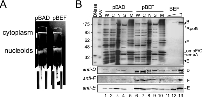
Copurification of MukBEF with nucleoids. Nucleoids were isolated from DH5α cells that carried either the vector (pBAD) or the MukBEF-overproducing plasmid pBB03 (pBEF) 1 h after induction with arabinose. The isolated nucleoids were then separated into the scaffold and the membrane fractions as described in Materials and Methods. (A) Separation of the nucleoids from the cytoplasm using sucrose gradient centrifugation. Following centrifugation, the tubes were placed against a dark background and illuminated from the top. The cytoplasm and nucleoids were collected as indicated using wide-bore pipette tips. (B) SDS-PAGE analysis of isolated nucleoids. Whole-cell extract (W), cytoplasm (C), nucleoids (N), scaffold (S), and membranes (M) were resolved on a 10% gel along with increasing amounts of purified MukBEF (BEF). The proteins were visualized by silver staining (top panel) or by immunoblotting (bottom panels). Lanes 1 to 3, 5 μg of total protein; lanes 6 to 8, 3 μg of protein. A total of 5 μg protein as in lane 3 was separated into the scaffold and membrane fractions and analyzed in lanes 4 and 5, respectively. Similarly, 3 μg of protein as in lane 8 was fractionated for lanes 9 and 10. Lane 13 contains purified MukBEF (2.2 pmol of MukB-His10, 1.6 pmol of MukF, and 1.6 pmol of MukE). Lanes 11 and 12 contain 100-fold and 10-fold dilutions of MukBEF, respectively, as in lane 13. Arrowheads mark positions of MukB (B), MukF (F), and MukE (E). Also marked are positions for RNA polymerase (RpoB) and the major outer membrane proteins OmpC and OmpA. RpoB was identified from the comparison with the previous studies (18, 19) based on its molecular mass (156 kDa) and the large abundance of the protein in the nucleoid fraction. Note that MukB is not the major protein in the scaffold fraction even after overproduction.

