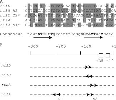
FIG. 7.
Comparison of HilC/HilD binding sites in the rtsA, hilC, hilD, and hilA promoters. (A) Alignment of the HilC/HilD binding sites defined by DNase I footprinting here and previously (37). The common positions are indicated by shading. The proposed consensus is shown in the bottom. The arrows indicate two direct repeats of CNATTNNT (shown in bold). Predominant bases (5/5 or 4/5) in the consensus sequence are indicated by uppercase letters, bases identical in 3/5 of the HilC/HilD binding sites are indicated by lowercase letters, and degenerate bases are marked by the letter N. The inverted positions of hilC C2 and hilA A1 sites in alignment compared to regions in hilD, rtsA promoters and hilA A2 site are marked by asterisks. (B) Positions and directions of the HilD/HilC consensus in the rtsA, hilC, hilD, and hilA promoters relative to the transcription start point, indicated as +1.

