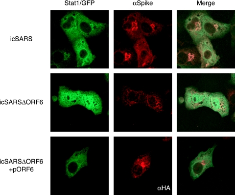
FIG. 2.
STAT1 localization during SARS infection. STAT1/GFP plasmid was transfected into Vero cells, and its localization was assayed after SARS infection. At 24 h posttransfection, cells were infected with either icSARS (top) or icSARSΔORF6 (middle) at an MOI of 3 for 12 h. Before fixation in 4% PFA, cells were treated with 100 IU of IFN-β for 1 h. The cells in the bottom panels were cotransfected with STAT1/GFP and HA-ORF6 prior to infection with icSARSΔORF6. Cells were then labeled with anti-SARS spike antibody to visualize the SARS-infected cells or anti-HA antibody (to visualize the transfected cells) and an Alexa 546-conjugated secondary antibody. Cells were visualized by using a confocal microscope.

