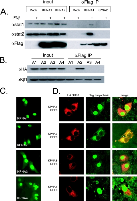
FIG. 3.
ORF6 affects localization of KPNA2. (A) 293 cells were transfected with Flag-tagged karyopherin plasmids. At 24 h posttransfection, half of the cells were treated with 100 IU of IFN-β for 30 min, and protein was harvested as described in Materials and Methods. Proteins were then immunoprecipitated (IP) with anti-Flag (αFlag) M2 antibodies and run on an SDS-PAGE gel before being transferred for Western blotting. Blots were probed with STAT1, STAT2, or Flag antibodies. + indicates treatment with 100 IU/ml of IFN-β. (B) Immunoprecipitations were performed on 293 cells cotransfected with each Flag-tagged karyopherin and HA-tagged ORF6. Lysates were prepared and immunoprecipitated as described in Materials and Methods. The top panel was probed with anti-HA antibody to visualize ORF6, and the bottom panel was probed with anti-KPNB1. A1, KPNA1; A2, KPNA2; A3, KPNA3; A4, KPNA4. (C) Vero cells were transfected with Flag-tagged KPNA plasmids. At 24 h posttransfection, cells were fixed and labeled with anti-Flag antibody. (D) Vero cells were cotransfected with Flag-tagged KPNA plasmids and HA-ORF6 plasmid. At 24 h posttransfection, cells were fixed and labeled with anti-Flag antibody and anti-HA antibody. Anti-Flag antibody was visualized with Alexa Fluor 488-conjugated secondary antibody, and anti-HA antibody was visualized with Alexa Fluor 546-conjugated secondary antibody. Note the overlapping localization of ORF6 and KPNA2.

