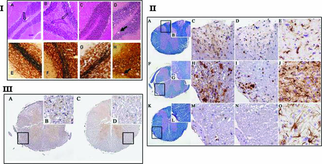
FIG. 4.
(I) H&E and Bielschowsky staining of cerebella from coinduced mice and controls. Infiltrates of mononuclear cells were detected in cerebella of all mice immunized with MOG. Axonal loss was also detected in scattered areas. There was no statistical difference in the numbers of infiltrates or the loss of axons in both EAE-induced and coinduced groups of mice. In scrapie-infected cerebellum, we could detect some degree of axonal loss and no cellular infiltrates. (II) Demyelination and inflammation in spinal cords. (A to E) EAE-induced animal. (A) Spinal cord shows small areas of demyelination after immunization with MOG (Luxol fast blue/PAS stain) (rectangle in panel A is enlarged in panel B). Original magnification, ×40 (A) or ×200 (B). (C) Scattered macrophages (MAC3 stain). Original magnification, ×400. (D) Lymphocytes (CD3 stain). Original magnification, ×400. (E) Moderate gliosis is visible in gray matter of the medulla oblongata (GFAP stain). Original magnification, ×400. (F to J) Coinduced animal. (F) Spinal cord shows more prominent and florid demyelination after PrPSc inoculation and MOG immunization (Luxol fast blue/PAS stain) (rectangle in panel F is enlarged in panel G). Original magnification, ×40 (F) or ×200 (G). (H) Numerous macrophages (MAC3 stain). Original magnification, ×400. (I) Lymphocytes (CD3 stain). Original magnification, ×400. (J) Profound gliosis is visible in gray matter of the medulla oblongata (GFAP stain). Original magnification, ×400. (K to O) Scrapie-incubating animal. (K) Spinal cord does not show any demyelination after PrPSc inoculation (Luxol fast blue/PAS stain) (rectangle in panel K is enlarged in panel L). Original magnification, ×40 (K) or ×200 (L). (M and N) No inflammation (MAC3 stain [M] and CD3 stain [N]). Original magnification, ×400. (O) Moderate gliosis is visible in gray matter of the medulla oblongata (GFAP stain). Original magnification, ×400. (III) PrPSc deposition in coinduced and scrapie-incubating mice. A spinal cord after PrPSc inoculation (homogenate, 10−2) and immunization with MOG shows PrPSc deposition in gray matter (A) and in the demyelinated white matter (B) (rectangle in panel A is enlarged in panel B) (6H4 stain). Original magnification, ×40 (A) or ×600 (B). A spinal cord after PrPSc inoculation (diluted 10−8) shows PrPSc deposition in gray matter (C) and unaffected white matter (D) (rectangle in panel C is enlarged in panel D) (6H4 stain). Original magnification, ×40 (C) or ×600 (D).

