FIG. 6.
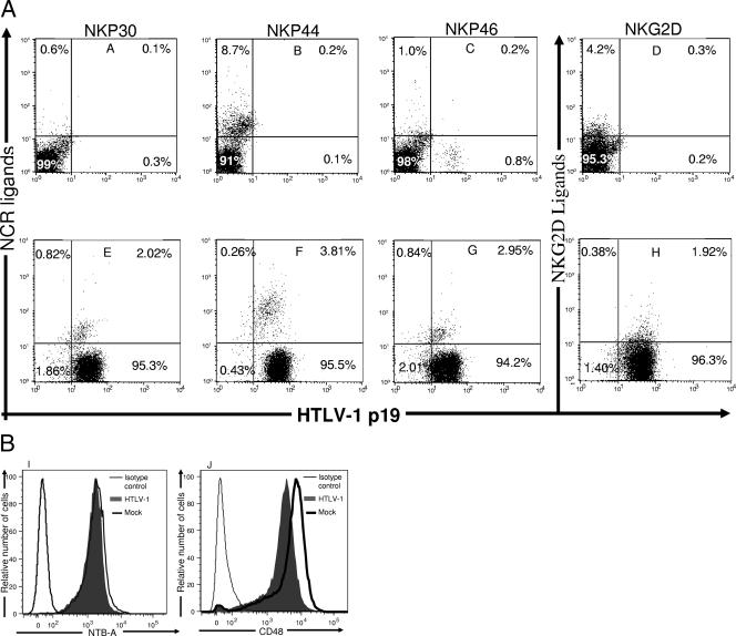
Expression of ligands for NCR, NKG2D, 2B4, and NTB-A on HTLV-1-infected primary CD4+ T cells. Mock-infected (A to D) and HTLV-1-infected (E to H) CD4+ T cells were stained with recombinant human NKp30 and IgG Fc fusion proteins (A and E), human NKp44 and IgG Fc fusion proteins (B and F), human NKp56 and IgG Fc fusion proteins (C and G), and human NKG2D and IgG Fc fusion proteins (D and H). Cells were then permeabilized, fixed, and stained with anti-HTLV-1 p19 capsid antibody. Numbers represent the percentage of cells in each quadrant. Mock-infected and HTLV-1-infected CD4+ T cells were stained with mouse anti-human NTB-A (I) and CD48 (J) antibody. Cells were then permeabilized, fixed, and stained with anti-HTLV-1 p19 capsid antibody. The extent of CD48 and NTB-A expressed on 104 infected cells (filled gray line) and 104 mock-infected CD4+ T cells (thick black line) were determined. Negative controls (thin black line) consisted of secondary antibody in the absence of primary antibody.

