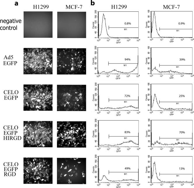
FIG. 4.
Ad-directed EGFP expression in human H1299 and MCF-7 cell lines. (a) Fluorescence microscopy of H1299 and MCF-7 cells following infection with Ad5-EGFP and with CELO-EGFP, CELOEGFP-RGD, and CELOEGFP-HIRGD viruses at an MOI of 10,000 vp/cell for 48 h (Olympus IX-71 microscope; magnification, ×350; Olympus, Germany). Representative fields are presented. (b) Quantitative assessment of Ad vector-mediated EGFP expression. The results of flow cytometric analysis of H1299 and MCF-7 cells infected with the Ad5 and CELO vectors indicated in panel a are shown. The cells were infected for 48 h at an MOI of 10,000 vp/cell. The percentage of EGFP-positive cells as defined by gate M1 is given in each panel.

