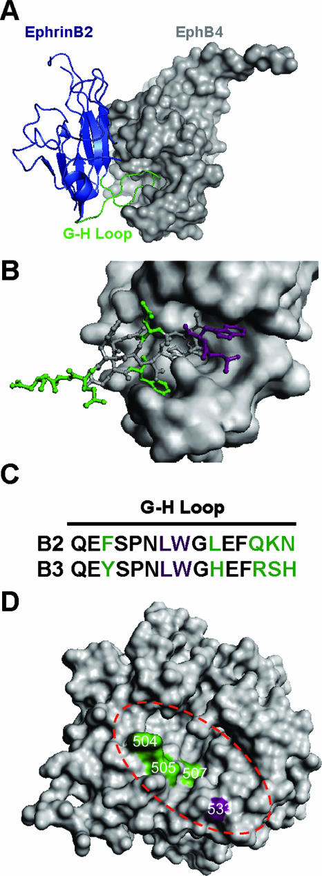
FIG. 7.
NiV-G model and proposed site of ephrinB2 and -B3 interaction in comparison to the crystal structure of the ephrinB2-EphB4 complex. (A) The crystal structure of an ephrinB2 monomer (blue cartoon representation) in complex with its cognate receptor, EphB4 (surface representation), indicates that the G-H loop of ephrinB2 (green) inserts into a hydrophobic canyon of EphB4 (11). (B) Detailed view of the G-H loop of ephrinB2 (stick representation) that makes contact with the binding cleft of EphB4 (surface representation). (C) Alignment of the G-H loop residues of ephrinB2 and -B3. Color-coded residues correspond to those indicated in panel B. The purple residues are critical for ephrinB2 and -B3 binding to NiV-G, and the green residues indicate the differences between ephrinB2 and -B3 in the G-H loop. (D) Structural model of the NiV-G protein globular domain described by Guillaume et al. (20), displayed as a top view with surface representation. Green residues (W504, E505, and V507) localize to the top surface of the NiV-G model and represent residues involved in distinct ephrinB3 binding. The purple residue (E533) has overlapping ephrinB2 and -B3 binding properties. The dashed red oval highlights a predicted binding cleft or canyon in the NiV-G model. All images were created using PyMol v0.99 (DeLano Scientific LLC, Palo Alto, CA).

