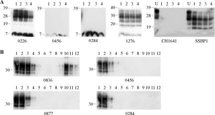
FIG. 2.
Analysis of ovine cerebellum samples from clinical scrapie-affected animals. (A) Samples were digested by the addition of 150 μg/ml thermolysin every hour for a total of 8 h. Resulting digests after 1, 4, 6, and 8 h of digestion are shown in lanes 1 to 4, respectively. For experimental scrapie strains SSBP1 and CH1641, undigested controls (U) are also shown. Digested brain homogenate was loaded at 3.3 μl of 10% (wt/vol) per lane, and undigested samples were loaded at 3.3 μl of 2% (wt/vol) per lane. PrP was detected on Western blots with monoclonal antibody P4. (B) Samples (200 μl of 10% [wt/vol] cerebellum homogenate) were resolved through a 10 to 60% sucrose step gradient and 12 0.42-ml fractions were collected. Each fraction (18 μl) was analyzed by Western blotting (lanes 1 to 12 for fractions taken from the top to bottom of the sucrose gradients), and PrP was detected with monoclonal antibody SAF32. For all blots, animal reference numbers and molecular mass markers (kilodaltons) are indicated. All samples in panel A are from clinical scrapie-affected animals; in panel B, animals 0836, 0456, and 0284 are clinical scrapie cases, and animal 0877 is a healthy control.

