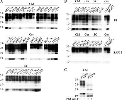
FIG. 3.
C2 PrP species within the CNS of TSE-affected sheep. Samples from clinical scrapie- or BSE-affected sheep of various PrP genotypes were analyzed. Brain homogenates (3.3 μl of 2% [wt/vol]) were analyzed directly (panels A and B) or were deglycosylated before analysis (panel C as indicated). PrP was detected on Western blots with P4 (all panels) or SAF32 (panel B as indicated). Included were caudal medulla (CM), spinal cord (SC), and cerebellum (Cer) samples. Material from field isolates of scrapie (from animals 0923, 0615, 0456, 0284, 0455, 0925, 0836, 0226, 1276, 1563, and 1275) as well as experimental scrapie (from strains CH1641 and SSBP1), experimental BSE (from animals 0392, 1693, and 0654), and samples from a healthy control animal (C1) are shown. Prolonged exposures of the blots are shown in the bottom panels, highlighting the presence of unglycosylated C2 fragment. For all blots, animal reference numbers and molecular mass markers (kilodaltons) are indicated. −, absence of PNGase F treatment; +, presence of PNGase F treatment.

