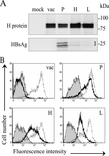
FIG. 2.
HBsAg expression by vectored MV. (A) Immunoprecipitation of proteins produced by Vero/hSLAM cells infected with MVvac2 (vac), MVvac2(HBsAg)P (P), MVvac2(HBsAg)H (H), and MVvac2(HBsAg)L (L). Mock, mock-infected cells. Proteins were labeled with [35S]methionine 20 to 24 h postinfection and precipitated either with an H-specific serum (top) or an HBsAg-specific serum (bottom). The positions of molecular mass standards are indicated on the right. Arrowheads indicate two weak HBsAg signals. (B) Fluorescence-activated cell sorter analysis of HBsAg and MV N protein expression in infected cells. Thick lines, HBsAg expression; thin lines, N expression; shaded area, negative control without primary antibody.

