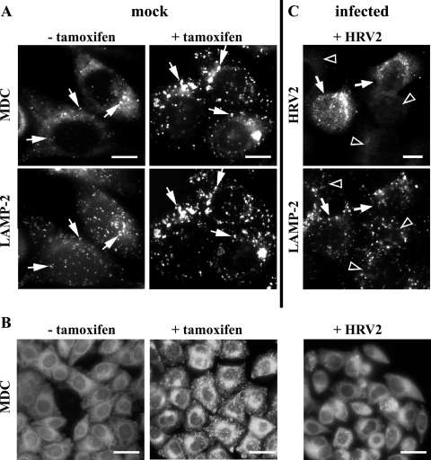
FIG. 1.
Visualization of autophagosomes by MDC and LAMP-2. (A) Cells were incubated with (+) or without (−) 1 μM tamoxifen for 48 h and stained with MDC and anti-LAMP-2 antibody. A few LAMP-2-positive vesicles (arrows) were also stained with MDC in the absence of tamoxifen. However, large MDC- and LAMP-2-positive compartments are induced in all cells by the drug (arrows). (B) Control cells with or without tamoxifen and cells infected with HRV2 at 300 TCID50/cell were stained with MDC. Infected cells lacked the typical tamoxifen-induced MDC staining (right). (C) Infected cells were stained with anti-HRV2 and anti-LAMP-2 antibodies followed by Alexa 568 and Alexa 488-conjugated secondary antibodies. No alteration in LAMP-2-positive compartments was observed in cells actively replicating virus (arrows). Arrowheads, noninfected cells. Bars, 10 μm (A, C) and 30 μm (B).

