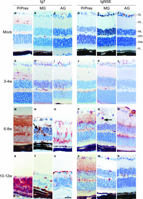
FIG. 3.
Immunohistochemical analysis of PrP-res, microglia, and activated astroglia in scrapie-infected tg7 and tgNSE retinas. Representative sections from uninfected (mock) or scrapie-infected mice sacrificed at 3 to 4 weeks (w), 6 to 8 weeks, or 10 to 12 weeks (clinical) p.i. are shown. Immunohistochemical staining was performed with anti-3F4 antibody (PrP-res), anti-Iba1 antibody for microglia (MG), or anti-GFAP antibody for activated astroglia (AG), as described in Materials and Methods. Compare activated microglia with thickened processes (arrow) in tg7 mice (N) to microglial cells with thin processes (arrow) in tgNSE mice (Q). Retinal layers are indicated as follows: PL, photoreceptor layer; ONL, outer nuclear layer; OPL, outer plexiform layer; INL, inner nuclear layer; IPL, inner plexiform layer; GL, ganglion layer. Scale bar, 20 μm (in panel U). Original magnification, ×40.

