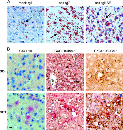
FIG. 8.
(A) Comparison of Iba1-positive microglia in the thalamus of scrapie-infected (sc+) tg7 and tgNSE mice at the time of clinical disease versus an aged-matched tg7 mock-infected control. Immunohistochemical staining was performed with anti-Iba1 antibody as described in Materials and Methods. The arrows indicate fine microglial processes in the mock-infected brain compared to thickened processes and more numerous cells in the infected brain. Scale bar, 20 μm. (B) In situ hybridization and immunohistochemical analysis of CXCL10 expression in astrocytes of mock-infected (sc−) or scrapie-infected (sc+) tg7 mice. Representative sections from the superior colliculus were hybridized with digoxigenin-labeled antisense RNA for CXCL10 and developed using Fast Red stain (bright pink around blue nuclei). The sections were then incubated with anti-Iba1 or anti-GFAP and developed with diaminobenzidine (brown/black). All sections were counterstained with hematoxylin. The blue arrows indicate colocalization of CXCL10 expression with anti-GFAP stain. This colocalization was not seen with anti-Iba1. Scale bar = 20 μm. Original magnification, ×40.

