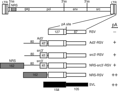
FIG. 1.
Schematic of in vitro polyadenylation substrates. At the top is a diagram of the RSV provirus showing the long terminal repeats (LTR); 5′ ss; NRS (shaded); gag, pol, env, and src genes; and 3′ ss. Below is an expansion of the 3′ LTR and a schematic of the RSV substrates, which include the entire region downstream of the poly(A) site (87 nt) (thin open box) and 127 nt of upstream sequence (wide open box). Ad3′-RSV has 47 nt of Ad 3′ exon (lightly shaded box) with 80 nt of upstream intron (thin line). src3′-RSV has 41 nt of the src 3′ exon (shaded box) with 80 nt of upstream RSV intron. NRS-src3′-RSV and NRS-RSV have the 162-nt NRS BBΔ76 fragment (dark shaded box) inserted upstream of the RSV poly(A) signal, with or without the src 3′ ss region. The positive control SVL substrate (black boxes) contains 137 nt of upstream and 105 nt of downstream sequence relative to the SVL poly(A) site. A summary of polyadenylation activity is indicated at the far right (−, activity less than 10% of that of SVL; +, activity of up to 40% of that of SVL; ++, activity of 40% or more of that of SVL).

