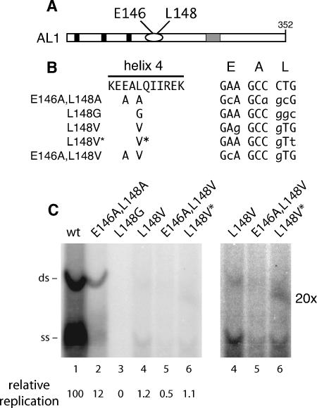
FIG. 1.
L148 mutants are impaired for TGMV AL1 replication. (A) Schematic of the TGMV AL1 protein. Solid boxes mark the locations of the three motifs conserved among rolling-circle replication initiator proteins, the oval indicates a predicted pair of α-helices, and the stippled box shows the location of the ATP binding motif. Helix 4 residues (E146 and L148) that were mutated are indicated. (B) The sequence between TGMV AL1 amino acids 144 and 154 (helix 4) is shown. E146 and L148 substitutions are shown for the five AL1 mutants below the sequence. The codons specifying residues E, A, and L of helix 4 and the mutations introduced into the three codons are shown on the right (modified nucleotides are indicated by lowercase type). Mutants L148V and L148V* differ only in the third position of the codon. (C) Replication of TGMV AL1 mutants was analyzed in tobacco protoplasts by agarose gel blot hybridization. Lanes 1 to 6 are transfections with TGMV A replicons with either wild-type (wt) (lane 1) or mutant AL1 genes corresponding to E146A L148A (lane 2), L148G (lane 3), L148V (lane 4), E146A L148V (lane 5), and L148V* (lane 6). The positions of double-stranded (ds) and single-stranded (ss) forms of TGMV A DNA are marked on the left. An overexposed image (magnification, ×20) of lanes 4 to 6 is shown on the right. The levels of replication of the different mutants relative to wild-type TGMV (100) are indicated at the bottom.

