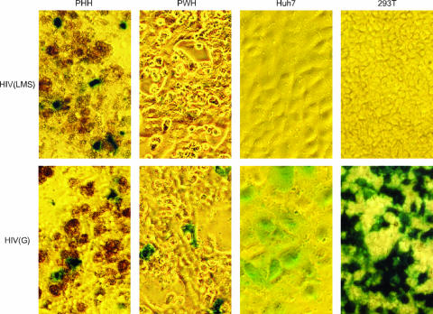
FIG. 2.
Light microscopy revealed infection of different cell types by HIV pseudotypes. HIV(LMS) or HIV(G) was used to infect PHH, PWH, Huh7 cells, or 293T cells. After 3 days, the cells were fixed and stained with X-gal. Infected cells were thus detected by blue staining. A brown signal was associated with some nonviable PHH.

