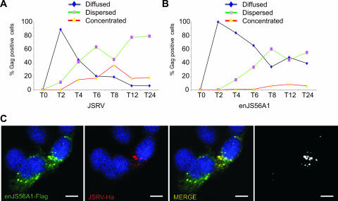
FIG. 1.
Kinetics of JSRV/enJS56A1 Gag staining pattern. (A and B) Quantification of JSRV and enJS56A1 Gag staining pattern in confocal microscopy of HeLa cells at various time points posttransfection. The number of cells in which Gag accumulation was observed diffused, dispersed, and concentrated was counted. Approximately 100 cells were evaluated at each time point. Graphs represent the average of two independent experiments. (C) HeLa cells expressing JSRVHA-MA and enJS56A1Flag-MA were fixed at the 4-h time point and analyzed by confocal microscopy using antibodies to the Flag (green) and Ha (red) epitopes and appropriate secondary conjugated antibodies. Scale bars, 10 μm.

