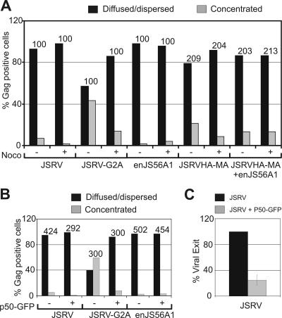
FIG. 3.
The targeting of the JSRV Gag to the centrosomal region is dependent on dynein and intact microtubule network. (A) Quantification of JSRV, JSRV-G2A, enJS56A1, and JSRVHA-MA Gag staining patterns by confocal microscopy in HeLa cells after treatment with nocodazole or with the solvent dimethyl sulfoxide used as a negative control. Gag proteins were detected by using antibodies to JSRV MA or HA. The graph represents the number of cells in which Gag staining was observed concentrated as opposed to diffused or dispersed. The number of Gag-positive cells counted in each experiment is indicated above each bar. Note that the use of anti-HA antibodies, compared to polyclonal anti-JSRV-MA serum, results normally in a relatively higher percentage of cells with concentrated Gag staining and a lower percentage of cells displaying diffuse Gag. These differences are probably due to the different sensitivity of the two antisera. (B) The effect of p50-GFP on Gag staining pattern was evaluated as in panel A. The number of Gag-positive cells counted in each experiment is indicated above each bar. Note that only cells double positive for Gag and p50-GFP were counted. (C) Graph representing the quantification of three independent SDS-PAGE and Western blotting experiments of viral particles released in the supernatant of 293T cells transfected with a JSRV expression plasmid in the presence or absence of p50-GFP. Filters were incubated with an antiserum to the JSRV CA. Signals were quantified by chemifluorescence as described in Materials and Methods. Bars indicate the standard errors.

