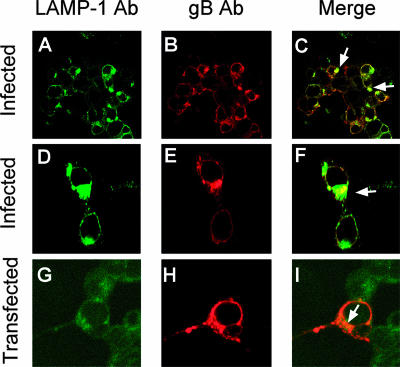
FIG. 4.
gB accumulates in part at MVB membranes in HSV-infected and -transfected cells. (A to F) 293T cells were infected with HSV-1 (10 PFU/cell). Twenty-four hours later, the cells were stained with MAb to gB (Virusys) and PAb to LAMP-1 and analyzed by confocal microscopy at a ×63 magnification objective. (G to I) 293T cells were transfected with a construct expressing gB (pgBwt-MTS). Fourty-eight hours later, the cells were stained with MAb to gB (Virusys) and a PAb to LAMP-1 and analyzed by confocal microscopy. The arrows point to colocalization spots. (A to C) Low magnification; (D to F) high magnification.

