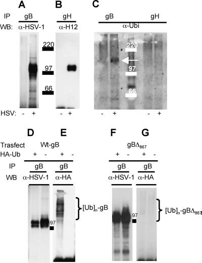
FIG. 6.
HSV-1 gB is ubiquitinated in infected and in transfected cells. (A to C) Electrophoretic and Western blotting analyses of gB and gH immunoprecipitated (IP) from lysates of HSV-1-infected (+) or uninfected (−) 293T cells by means of MAb H1817 to gB or MAb 53S to gH. The immunoprecipitated proteins were separated by SDS-PAGE, followed by Western blotting (WB) with the indicated antibodies (α-HSV-1, PAb to the major HSV-1 glycoproteins; α-H12, MAb to gH antibody; α-Ubi, PAb to ubiquitin). (C) The arrow points to the band of ubiquitinated gB. Numbers identify the electrophoretic mobilities of the molecular mass markers. (D to G) 293T cells were cotransfected with a construct expressing wt gB (pgBwt-MTS) (D, E) or a truncated form of gB (gBΔ867) (F, G). Cells were also transfected with a construct encoding HA-tagged wt ubiquitin (HA-Ub) or the corresponding pBJ5 empty vector, indicated as HA-Ub + or HA-Ub −, respectively. Forty-eight hours posttransfection, gB was immunoprecipitated with MAb H1817. The separated proteins were analyzed by Western blotting with the antibodies listed above. Braces identify polyubiquitinated forms of gB. 97 indicates the electrophoretic mobility of the molecular mass marker.

