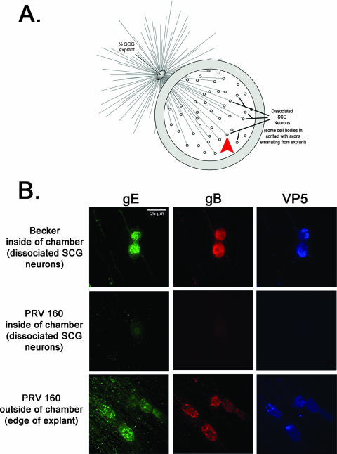
FIG. 1.
Us9 is essential for transneuronal spread in vitro. (A) Diagram illustrating the isolator chamber system in which one-half of an SCG explant is plated on Aclar and allowed to extend neurites. A Teflon chamber ring is then placed on top of preformed axons, capturing a subpopulation of axon ends. Dissociated SCG neurons are then plated inside the chamber, where they mature and form synaptic connections with axons emanating from the explant. Dissociated SCG neurons inside the chamber are equivalent to one-fourth of an SCG ganglia, which is approximately 5,000 neurons. All chambers were prepared in duplicate. The red arrowhead denotes where the images in panel B were taken in reference to the SCG explant. (B) Explants were infected on the outside of the chamber with wild-type PRV Becker or PRV 160 (Us9-null). At 24 h postinfection, both the explant and the dissociated neurons inside the chamber were fixed and labeled with antibodies that recognize the viral glycoproteins gE and gB, as well as the major capsid protein VP5. Postsynaptic neurons in contact with the Becker-infected SCG explant stained for viral glycoprotein and capsid proteins (top row). No labeling of viral antigen was detected in postsynaptic neurons in contact with the explant infected with PRV 160 (middle row), although the explant itself showed extensive infection (bottom row).

