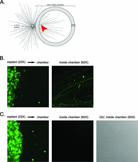
FIG. 3.
GFP-tagged capsids do not enter the axon in the absence of Us9. (A) A chamber ring was placed on top of preformed axons emanating from the SCG explant to physically separate the site of infection from the site of imaging. No dissociated SCG neurons were plated inside the chamber. The arrowhead highlights the region where images were taken at high magnification within the chamber. (B and C) Explants were infected for 24 h with PRV GS443 (GFP-VP26) (B) or PRV 368 (GFP-VP26, Us9 null) (C) and fixed with 4% paraformaldehyde. Direct fluorescence of explants outside the chamber (×20 magnification) and capsids inside the chamber (×60 magnification) was visualized by using spinning-disk confocal microscopy.

