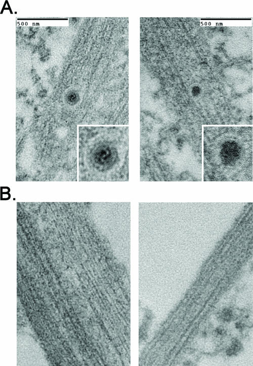
FIG. 4.
Axons are devoid of enveloped virus particles during a Us9-null infection. Explants on the outside of the chamber were infected for 24 h with PRV Becker or PRV 160 (Us9-null), and axons inside the chamber were visualized by transmission electron microscopy. Samples were examined in duplicate. A 1-mm square with high axonal density inside the chamber was selected for sectioning by the ultramicrotome. Six serial sections (70 nm apart) were scrutinized for the presence of virus particles. (A) Enveloped virus particles were detected in the distal-axon of Becker-infected explants but were not present in explants infected with PRV 160 (B).

