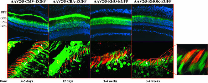
FIG. 2.
Subretinal injections of AAV2/5-EGFP vectors with various promoters into adult C57BL/6 mice. The images in the top row depict fluorescence microscopy evaluation of EGFP expression 4 weeks after subretinal injections of AAV2/5 encoding EGFP from the CMV, CBA, RHO, and RHOK promoters. Abbreviations: ONL, outer nuclear layer; INL, inner nuclear layer; GCL, ganglion cell layer. The images in the bottom row show staining with PNA-lectin (red label) on retinal sections of animals injected with AAV2/5-CMV-EGFP, AAV2/5-CBA-EGFP, AAV2/5-RHO-EGFP, and AAV2/5-RHOK-EGFP. Colocalization of EGFP expression and PNA staining is indicated by the arrows. Confocal microscope magnification, ×63. The selected field shows the colocalization of cone sheathes (PNA, red label) and EGFP expression (green label) at increased magnification. The magnified portion depicts the onset of gene expression as assessed by indirect ophthalmoscopy.

