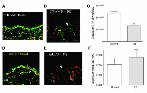
Figure 1. PS downregulates epidermal AMP expression.
Normal hairless mice (n = 3 each for immunohistochemistry and RT-PCR studies; 3–4 replications for each experiment in these and subsequent experiments) were exposed to insomnia- and crowding-induced PS (B and E) for 72 h, while littermate controls (A and D) were not stressed. Frozen sections (8 μm) were stained with primary antibodies to CRAMP and mBD3 and processed as described in Methods (for controls, see Supplemental Figure 1). Throughout the figures, white arrows indicate normal levels of positive immunostaining (green); white arrowheads indicate reduced staining; asterisks indicate positive immunostaining of pilosebaceous follicles; and “d” and “e” indicate dermis and epidermis, respectively. (C and F) mRNA was extracted from PS and control mouse epidermis, followed by quantitative RT-PCR (see Methods). Normalization in this and all subsequent studies was to 18S mRNA, with 2–3 replicates per sample (n = 3 per cohort). Scale bars: 50 μm. *P = 0.007.

